- For Appointment:
- +91 63590 11009
- Chat on Whatsapp

Linitis plastica is a stomach cancer that spreads inside the wall of the stomach. The wall turns thick and stiff, while the inner lining can look almost normal. This page is about the operation: the short keyhole look that comes first, the removal of the whole stomach, the margins, where the robot helps and where open hands are safer. Apollo Hospital, Bhat, Gandhinagar · Gota OPD, Gota, Ahmedabad.
Last updated: 18-Oct-2026
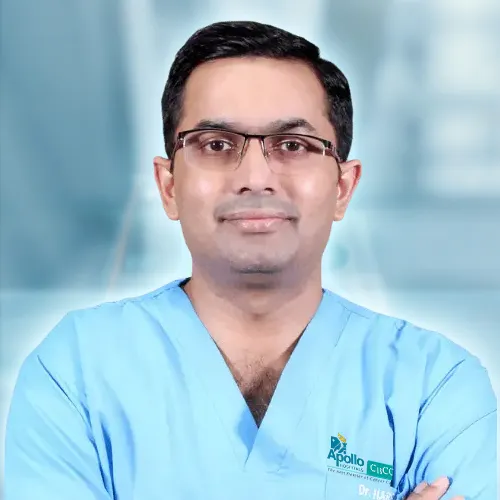
Dr Harsh Shah
MS, MCh (Surgical Gastroenterology), DrNB (Surgical Gastroenterology)
Registered with Gujarat Medical Council, Reg. No. G-38738
Cancer Surgeon (GI)
Dr. Jivraj Mehta Hospital, Vasna, Ahmedabad · 18 years of experience
★★★★★ Google: 5.0 from 25 reviews (Dr. Jivraj Mehta Hospital, Vasna, Ahmedabad)
Dr Harsh Shah. Two higher degrees in GI surgery and 18 years of experience in GI and cancer surgery, including robotic stomach surgery. Every stomach cancer is discussed by a tumour board of surgeons, cancer doctors, scan doctors and pathologists before treatment. Dr Harsh Shah performs the operation himself, at the robot console or at the open table.
In short
Linitis plastica needs a keyhole look inside the abdomen before any big operation. Hidden spread on the lining is common, and no scan reliably shows it.
When an operation is advised, the whole stomach is removed with its lymph nodes. The cut edges must be checked under the microscope during the operation.
The robot helps with the deep lymph node work and the high join. But it cannot feel the stiff wall, so open surgery is often the safer choice here.
In linitis plastica the cancer cells spread through the layers of the stomach wall. A biopsy from the surface can come back clear, even when the wall is full of cancer.
Many families first hear the word after a confusing time. Meals feel too big. Weight falls. A scan shows a thick stomach wall. Yet the first biopsy says no cancer. That is the nature of this disease. The cells grow under the inner lining, not on top of it.
This page starts after that stage. Your diagnosis has been confirmed, and the tumour board has seen your reports. We do not repeat the tests here. Your doctors will already have them.
What matters now is how this kind of cancer changes the operation. Four things set it apart from other stomach cancers:
Before any stomach is removed, a short keyhole operation looks at the lining of the abdomen and collects washings. It is the most useful half hour in this whole plan.
Our protocol asks for this look, called a staging laparoscopy, before any curative plan for a stomach cancer of this kind. Our technique notes name linitis plastica as the cancer most likely to have hidden spread. So the look is never skipped here.
This first look is done with keyhole instruments, not with the robot. A camera goes in near the navel. One or two small extra ports are added. It is a short procedure under general anaesthesia.
The look may be done on its own, before chemotherapy starts. It may also be the first step on the day of the main operation. Your consent covers one hard possibility: if spread is found, the big operation is not done.
For most patients who can have an operation, chemotherapy comes first. The operation follows a few weeks after the last cycle, and more chemotherapy follows recovery.
Our protocol uses chemotherapy on both sides of the operation for stomach cancers that are not very early. It aims to shrink the cancer and treat cells that may already be travelling. The cancer doctor plans the drugs and the number of cycles with you.
Food is part of the treatment. Linitis plastica makes the stomach small and stiff. Many people cannot eat a normal meal. A dietitian sees you early. If you cannot keep up your weight, a thin feeding tube may be placed into the small bowel. It feeds you through the chemotherapy months, so you reach the operation strong.
Before the operation, the team scans you again. Then the tumour board confirms that removal is still the plan.
Our protocol names linitis plastica as a reason for total gastrectomy. Because the cancer runs through the wall, keeping part of the stomach would leave cancer behind.
In some stomach cancers, the lower part of the stomach can be removed and the upper part kept. That is not the case here. The cancer spreads along the wall, often far beyond what is seen. So the whole stomach is removed, with the fatty apron that hangs from it.
The lymph nodes around the stomach come out with it. This is called a D2 dissection. The nodes run along the main arteries above the pancreas and towards the spleen. The laboratory counts them and reports each group.
The spleen is usually kept. Our protocol does not remove the spleen or part of the pancreas as a routine.
Then food needs a new path. The lower end of the food pipe is joined to a loop of small bowel. This is called a Roux-en-Y join. Food then passes from the food pipe straight into the small bowel.
The hardest question in this operation is where to cut. Our protocol asks for wider margins than in other stomach cancers, and a microscope check when in doubt.
The cancer can extend beyond the part of the wall that looks or feels abnormal. If a cut edge contains cancer, it may return at the join. So the plan for the edges starts before the operation.
This is the step where open hands give the surgeon extra information. We explain that below.
The robot gives a steady, magnified 3D view and instruments that bend like a wrist. Its gain is in the deep lymph node work and in the join high under the diaphragm.
A total gastrectomy has two hard places. The first is the lymph nodes above the pancreas and towards the spleen. The second is the join between the food pipe and the bowel, made high up at the diaphragm.
Above the pancreas, nodes sit on and around the main arteries. With straight keyhole tools, the surgeon often has to press on the pancreas to reach them. Pressure on the pancreas can cause a leak of pancreatic juice. The robot’s wristed tools lift the nodes away instead. They peel the tissue off the pancreas without pushing on it.
Our technique notes describe two sealing instruments used together, one in each of the surgeon’s hands. They seal small vessels with little heat spread. That helps near the pancreas and the spleen’s vessels.
High at the diaphragm, space is tight. Wristed tools help the surgeon free the food pipe and make or check the join. A dye given into a vein can show on the robot’s screen whether the join has a good blood supply.
What does not change: the operation itself. The same stomach, the same nodes and the same margins are removed. Our notes say the robot does not change the cancer operation. It changes how the hard parts are done.
A robot has no sense of touch. When linitis plastica runs through much of the stomach, our technique notes make open surgery the default, so the surgeon can feel the edge.
In most stomach cancers, the edge is seen. In linitis plastica, it is often felt. The diseased wall is firm and thick. Healthy wall is soft. In open surgery, the surgeon’s fingers can trace that change along the stomach and the food pipe. The robot shows a sharper picture, but it cannot report stiffness.
Our protocol also says that for a total gastrectomy, or a bulky tumour, the approach is open unless the surgeon judges otherwise. So a robotic total gastrectomy for linitis plastica is offered only to selected patients.
| Your situation | Usual approach | Why |
|---|---|---|
| Clean first look, chemotherapy done, edge clearly seen and marked. | Robotic total gastrectomy can be considered. | The margin can be judged by eye, marks and frozen section. |
| Disease running through much of the stomach wall. | Open. | Touch helps find where the stiff wall ends. |
| Cancer growing into the colon, pancreas or spleen. | Open. | Removing other organs together is safer open. |
| Many past operations in the upper abdomen. | Often open. | Scar tissue hides safe planes. |
| Heart or lung disease that cannot take a long keyhole operation. | Open considered. | Gas pressure in the abdomen and a longer operation add strain. |
| Edge near the food pipe, nodes above the pancreas, no other organ involved. | Robotic or keyhole, in experienced hands. | Wristed tools help in the deep, tight areas. |
This is not a step back. Open surgery removes the same organ and nodes. For some people with this cancer, it is the more complete operation.
All three remove the whole stomach and the same lymph nodes. They differ in the cut, the view, the sense of touch, blood loss and how soon you are back on your feet.
| Open | Keyhole | Robotic | |
|---|---|---|---|
| The cut. | A long cut on the upper abdomen. | Small ports and one small cut. | Small ports and one small cut. |
| The view. | Direct. | Magnified, flat. | Magnified, 3D, held steady. |
| Feeling the stiff wall. | Yes, by hand. | No. | No. |
| Finding the margin. | Eye, touch and frozen section. | Eye, marks and frozen section. | Eye, marks and frozen section. |
| Nodes above the pancreas and near the spleen. | By hand. | Straight tools, often pressing on the pancreas. | Wristed tools lift the nodes off the pancreas. |
| The join high at the diaphragm. | Standard. | Hard in tight space. | Wristed tools help. |
| Lymph node standard. | The same. | The same. | The same. |
| Blood loss. | More. | Less. | Less. |
| Hospital stay. | Longest. | Shorter. | Shorter. |
| Return to work. | Later. | Sooner. | Sooner. |
| Switching to open during the operation. | Not needed. | More often. | Less often. |
| Cost. | Lowest. | Close to open. | Higher, for robotic tools. |
These are comparisons in words. Most research on robotic stomach surgery comes from early cancers, not from linitis plastica. The figures for your own operation are discussed at the consent visit.
You lie on your back with the head end raised. Small ports go across the upper abdomen, and the robot’s arms are attached once the first look is clear.
Sometimes the most useful decision is not to remove the stomach. Spread, or a body too weak for a long operation, makes medicines and support the better path.
Removing the whole stomach is a large operation. It is worth it only when it can remove all the cancer. Our protocol is clear about the times it cannot:
| Situation | Why not operate | What is offered instead |
|---|---|---|
| Cancer seen on the lining of the abdomen. | The operation cannot remove spread that is everywhere. | Medicines chosen by tests on the tumour tissue. |
| Cancer cells in the washings. | Our protocol treats it the same as spread. | Medicines first. Review by the tumour board. |
| Spread to the liver or distant organs. | Stage IV disease. | Medicines, nutrition and symptom support. |
| Too weak to be out of bed most of the day. | The body may not survive the strain. | Supportive care and a frank family discussion. |
| A narrowed stomach with severe vomiting, but spread present. | Removal will not help. | Feeding support or a stent, with a doctor’s same-day review. |
For every patient with spread, our protocol brings in nutrition and supportive care early. A second opinion is always welcome before a large operation, or before deciding against one.
Recovery follows a set plan from the first evening. You sit out of bed on the day of surgery, sip fluids, and walk the next day.
| When | What happens |
|---|---|
| Day of surgery. | Sitting out of bed a few hours after waking. Sips of water. Pain control with little or no morphine. |
| First day. | Liquid diet. Walking three times. The urine tube comes out. Feeding tube starts slowly, if you have one. |
| Second and third day. | Soft diet step by step. Drips are reduced. Pain tablets by mouth. |
| Fourth to sixth day. | Full soft diet. Any drain comes out. Checks for going home. |
Some signs are never watched overnight. A fast heartbeat, fever, new belly pain, or a change in drain fluid can mean a leak at the join. The team stops the diet, gives fluids into a vein and arranges a scan the same day. Call the office at once if any of these appear at home.
You go home when you have had no fever for a day, eat a soft diet, walk on your own and are comfortable on tablets. Eating changes for life.
Without a stomach, meals must be small and frequent, for life. Some people feel faint or sweaty after a large or sugary meal. A dietitian teaches you how to avoid this. Vitamin B12 is no longer absorbed, so injections continue for life. Iron is checked by blood tests.
Our protocol does not set a fixed return-to-work date. It depends on chemotherapy after surgery, your weight and your work. Desk work usually returns before heavy work. Your date is agreed at follow-up.
★★★★★
“My father has carcinoma of stomach ,Harsh sir did radical gastrectomy and he is recovering very nicely under Dr.Harsh's observation ,he listen each and every complain calmly and with patience and make patient very comfortable during treatment , during procedure and during stay at hospital. we are satisfied with the treatment and very confident about the fully recovery of disease as we are under treatment of Dr.Harsh & team.Thank you Dr.Harsh & his whole team and staff.”
tina parmar · Google review
★★★★★
“Excellent doctor for stomach cancer. We r very happy. Recommend for gastro cancer treatment in Ahmedabad”
Purshottam Meena · Google review
★★★★★
“We are much satisfied with robotic surgery done by Dr harsh shah. He is very humble in nature. Highly recommended for gastro cancer surgeon in Ahmedabad. Thank u so much sir.”
Jyotsnaben Kotwal · Google review
Quoted exactly as written on the centre’s Google listings. Two are about stomach cancer surgery and one about robotic surgery. None is presented as a case of linitis plastica.
A robotic operation costs more than an open one, because of the robotic tools. The keyhole look, a feeding tube or a longer stay also change the total.
The office prepares a written estimate once the operation is decided. Cashless admission is arranged at Apollo Hospital, Bhat when your policy allows it. Allow a day or two for robotic approval. Ask whether your policy covers robotic surgery, and up to what limit. Chemotherapy is planned and costed separately. You can also call the office on +91-63590-11009. Bring the policy papers, photo identity, all reports with scans on disc, and your medicine list.
Sometimes. If the first look is clean and the edge can be seen and marked, a robotic total gastrectomy can be considered. When the disease runs through much of the wall, open is usually safer.
No. It is a short keyhole look with a camera and one or two small ports. It comes before the big operation.
The cancer spreads along the wall, often beyond what is seen. Keeping part of the stomach would risk leaving cancer behind.
Our protocol treats this as spread. The stomach is not removed at that time. Medicines come first, and the tumour board reviews the plan.
Usually not. Our protocol keeps the spleen unless the cancer involves it.
Small meals, often, for life. A dietitian guides you. Vitamin B12 injections continue for life.
Dr. Jivraj Mehta Hospital, Dr Jivraj Mehta Marg, Vasna, Ahmedabad 380007
Consultation, review of scans and reports, second opinions, admission, 3D HD laparoscopic and open surgery, and follow-up. OPD Monday to Saturday, 9 am to 5 pm. Consultation fee ₹300.
Google: 5.0 · 25 reviews
+91-63555-64601 · +91-63590-11009 · Directions to Dr. Jivraj Mehta Hospital
Patients travel for stomach cancer surgery from across Gujarat, southern Rajasthan and western Madhya Pradesh.
Send the endoscopy and biopsy reports, the CT scan with its report, and any chemotherapy notes ahead of your appointment, so the consultation starts with them already read. Whether you need an operation, and whether it is robotic, keyhole or open, is decided at the consultation after examination, and by the tumour board. Call +91-63590-11009 WhatsApp the office
Swasth Parivar: every week, practical advice on recovery, diet and warning signs. No advertising. Stop any time.
Your number is used for these updates and for the clinic to reach you. It is never sold or passed on. To stop, reply STOP on WhatsApp.
