- For Appointment:
- +91 63590 11009
- Chat on Whatsapp


Most breast lumps are not cancer. The common causes are fibroadenomas, cysts and ordinary thickening that changes with the cycle.
A lump still deserves a proper look, because examination and a scan are the only reliable way to tell one kind from another. It is carried out by Dr Swapnil Sharma, MBBS, MS, FMAS, DrNB (Surgical Oncology), Consultant Surgical Oncologist, at Apollo Hospital International Ltd, Bhat, Gandhinagar.
Finding a lump is unsettling, and the first thing worth saying is that the odds are in your favour. Breast clinics see far more benign lumps than cancers. This page sets out what the usual causes are, how a lump is assessed, and when it makes sense to be seen.

| Cause | What it is | Who it is common in |
|---|---|---|
| Fibroadenoma | A smooth, firm, mobile lump of normal breast tissue that has grown as a discrete lump. Harmless. | Teens to thirties |
| Cyst | A pocket of fluid. Can appear quickly and can be tender. | Thirties to fifties |
| Cyclical thickening | Lumpy, ropy tissue that changes with the monthly cycle and settles after a period. | Any age before menopause |
| Fat necrosis | A firm area where bruised fatty tissue has healed, often after a knock or an operation. | Any age |
| Infection or abscess | A hot, red, painful area, most often while breastfeeding. | Any age |
| Cancer | Less common than the above, and treatable, particularly when it is found early. | Rises with age |
These are features that make a doctor want to look more carefully. They are not a diagnosis, and none of them on its own means cancer.
A lump that is hard rather than rubbery, or that feels fixed to the skin or the chest wall rather than moving freely. A lump that is getting larger over weeks. Dimpling or puckering of the skin over it. A nipple that has newly turned inwards, or blood-stained discharge from one nipple. A lump in the armpit. A lump in a man.
If one of these is present, arrange a visit rather than waiting to see what happens. If none is, a lump still present after one monthly cycle is the sensible point at which to have it looked at.
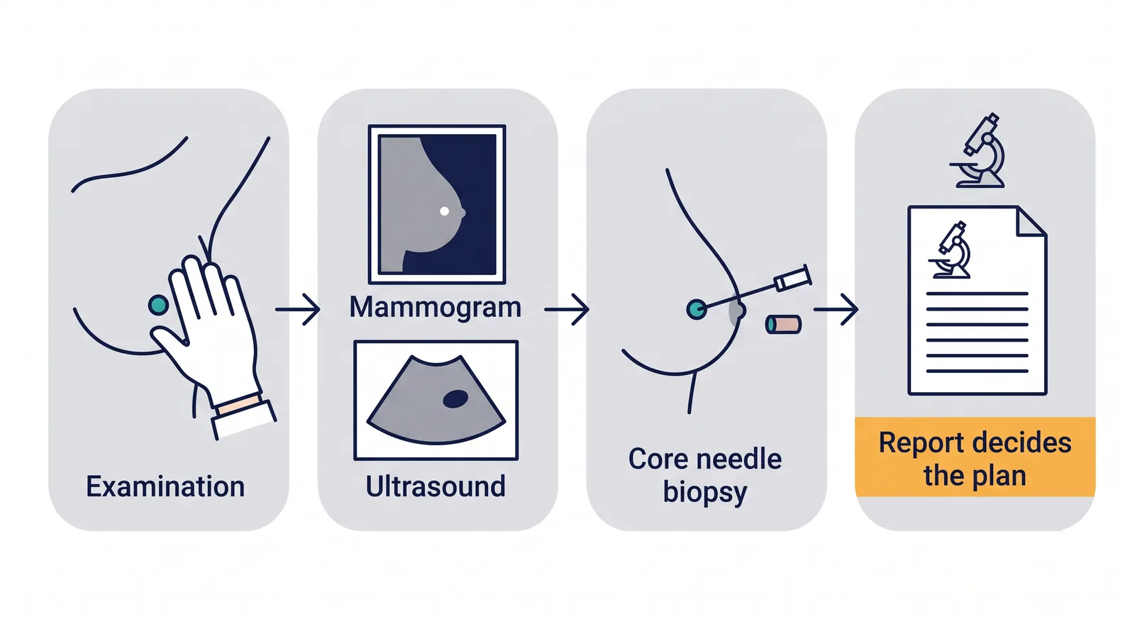
Examination, then a scan, then a needle sample only if the first two leave a question. Most women do not need the third step.
The assessment is deliberately stepwise, and it is usually finished in one visit.
Examination. Both breasts and both armpits. Size, texture, whether it moves, and whether the skin or nipple has changed.
Imaging. An ultrasound is the first test under about forty and for any lump that needs a closer look. Over forty a mammogram is usually added, because dense tissue thins with age and mammography reads the breast better then. Ultrasound tells a cyst from a solid lump immediately.
A sample, if needed. If imaging is clearly benign, no sample is taken. If there is a question, a core needle biopsy under local anaesthetic gives an answer in a few days. The fuller account is on the diagnosis page.
A cyst is drained with a fine needle. Small fibroadenomas are watched. Larger ones can be removed through a small cut.
If it is a cyst, draining it with a fine needle settles it, and often that is all that is ever needed. If it is a fibroadenoma, small ones are usually left alone and simply watched; larger ones, or ones that keep growing, can be removed through a small cut if you would rather not live with it.
If it is an infection, antibiotics treat it and an abscess is drained. If the sample does show cancer, the treatment routes are set out on the breast cancer surgery page, and most early breast cancer today is treated without removing the breast.
Being familiar with how your breasts normally feel is more useful than a formal monthly ritual. Breasts are naturally lumpy and that lumpiness changes through the cycle. What matters is noticing something that is new for you and that stays.
The easiest time to feel is a few days after a period ends, when the tissue is least swollen. After menopause any time is fine.
Usually not. Most breast lumps turn out to be benign, and that is true across all age groups. It is still worth having one looked at, because the only reliable way to tell one kind from another is to examine it and image it.
A fibroadenoma, which is a smooth rubbery lump common in younger women. A cyst, which is a pocket of fluid. Ordinary lumpy thickening that changes with the monthly cycle. An area of infection or inflammation. These are more common than cancer.
Pain is not a reliable guide in either direction. Benign lumps are often painful and cancers often cause no pain at all, but there are exceptions both ways. A lump is assessed on examination and imaging, not on whether it hurts.
There is no need to rush to a hospital the same day. A lump that is still there after one monthly cycle is worth having looked at without putting it off for months. If it is growing, or the skin or nipple has changed, arrange to be seen sooner.
An examination, then an ultrasound, and a mammogram as well if you are over about forty. If the picture is clear and benign, that is often the end of it. If there is any doubt, a needle sample settles it.
It is done with local anaesthetic and most women describe pressure rather than pain. It does not spread cancer. That worry is common and it is not supported by the evidence.
A surgeon or a breast clinic where the examination, the scan and the needle test can be done together, so you are not sent between places. Call +91 63555 64601 to fix a time at Apollo Hospital International Ltd, Bhat, Gandhinagar.
Send any scan you already have, then call to fix a time.
Dr Swapnil Sharma — MBBS, MS, FMAS, DrNB (Surgical Oncology)
Consultant Surgical Oncologist
Consultations and surgery: Apollo Hospital International Ltd, Bhat, Gandhinagar
Appointments: +91 63555 64601 · WhatsApp
Last updated: 24 September 2026. Reviewed by Dr Swapnil Sharma, MBBS, MS, FMAS, DrNB (Surgical Oncology), Consultant Surgical Oncologist.
This page is for information. It is not a substitute for an examination and a personal opinion.