- For Appointment:
- +91 63590 11009
- Chat on Whatsapp

Whichever level of the food pipe the cancer is in, removing it means working in two body cavities, and often a third short step in the neck. This page follows a robotic oesophagectomy phase by phase: what the robot changes in the chest, what it changes in the abdomen, and when the operation is done open or hybrid instead. Apollo Hospital, Bhat, Gandhinagar · Gota OPD, Ahmedabad.
Last updated: 8 October 2026
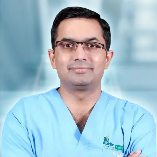
Shah’s Gastro, Cancer & Robotic Surgery Centre. Super-specialty training in surgical gastroenterology, two higher degrees in it, and 18 years of experience in GI and cancer surgery. Every food-pipe cancer is discussed at the tumour board before treatment begins, and Dr Harsh Shah performs the oesophagectomy himself.
An oesophagectomy removes the cancer-bearing food pipe with its lymph nodes and replaces it with a tube made from the stomach. It has a chest phase, an abdominal phase and, for many patients, a short neck step where the new food pipe is joined.
Done robotically or by keyhole, the chest and abdomen are operated through small ports instead of a long cut between the ribs and down the abdomen. The main gain is fewer lung complications, which are the most common serious problem after this operation.
In the chest the robot gives a steady, magnified view along the airway and the nerves to the voice box; in the abdomen it helps preserve the artery that will feed the new food pipe and checks its blood supply with fluorescence. Bulky tumours, scarring after radiotherapy or a difficult chest call for open or hybrid surgery.
A robotic or keyhole oesophagectomy suits a patient whose food-pipe cancer can be removed — with or without chemotherapy or chemoradiation first — and whose lungs and heart pass the tests done before any chest operation. It is the preferred approach here, because it reduces lung complications without compromising the cancer operation.
Fitness for this operation is measured, not assumed. Breathing tests including how well the lungs transfer oxygen, and a heart scan, are done before any chest phase; age alone does not decide it. Weeks of walking, breathing exercises and nutrition before surgery make a measurable difference, and a feeding tube is placed if swallowing has become too difficult to keep weight up.
Some patients are better served by open or hybrid surgery: a bulky tumour, a chest scarred and stiff after radiotherapy, previous surgery on the right side of the chest, severe lung disease, or a tumour pressing into the lining of the lung or the heart. A tumour growing into the airway, the aorta or the spine is not operated at all. An operation after full-dose chemoradiation, when cancer returns, is approached with particular caution and is often safer open.
Which treatment comes first for a particular tumour — operation, chemotherapy, chemoradiation, or chemoradiation alone — depends on the cancer type and its level, and is set out on the robotic oesophageal cancer surgery page and at the tumour board. This page assumes the decision to operate has been made, and explains what the robot changes in the operation.
The extent of the operation is the same. The robot changes how it is reached: in the chest, a magnified three-dimensional view along the airway and wristed instruments that clear the lymph nodes beside the nerves to the voice box; in the abdomen, fine control around the stomach’s blood supply and fluorescence imaging to confirm that the new food pipe is well perfused.
The food pipe runs through the back of the chest, between the airway, the aorta and the spine. The lymph nodes that matter most for squamous cancer lie along the windpipe, close to the thin nerves that move the vocal cords. Straight keyhole instruments reach these nodes from awkward angles. The robot’s wristed instruments and steady magnified view allow those nodes to be cleared while the nerves are seen and kept, which lowers the risk of a hoarse voice.
A robotic arm holds the lung out of the way for the whole chest phase, replacing a hand-held retractor. The large vein that arches over the food pipe, and the duct that carries fat-rich lymph up the chest, are identified and controlled in view.
In the abdomen, the stomach is freed while keeping the artery along its greater curve, because that artery will be the only blood supply of the new food pipe. A dye injected into a vein glows under the robot’s camera and shows how well the tip of the stomach tube is perfused; if the tip looks poor, it is trimmed before the join is made. There is no sense of touch through the instruments, so tumour edges are judged by sight and by the scans.
For the chest phase the patient lies turned partly face-down, with the right lung deflated and the other lung breathing for both. Robotic ports are placed between the ribs on the right side. For the abdominal phase the patient lies on the back, with ports across the upper abdomen and one arm holding the liver up. The neck step, when needed, is a short open cut on the left of the neck.
The half-prone position lets gravity draw the lung forward and gives a clear view of the back of the chest. It is chosen with conversion in mind: if the chest has to be opened, the patient must be turned, so the team decides the position for each patient before the operation, not during it.
Breathing on one lung for part of the operation is routine for the anaesthesia team, and is one reason the breathing tests before surgery matter. Positioning, padding and warming are completed before the robot is docked for each phase.
For a tumour in the middle or upper food pipe, the chest is usually done first, then the abdomen, then the neck, where the stomach tube is joined to the remaining food pipe. For a tumour in the lower food pipe or at the junction with the stomach, the abdomen is done first and the join is made inside the chest.
The margin above the tumour is planned in advance, and a frozen section is sent if there is any doubt about it.
The stomach, shaped into a tube, becomes the new food pipe. Its survival depends on one artery along its edge, so that artery is protected throughout and the blood supply at the tip is checked with fluorescence before the join. The join is the part of the operation most watched in the first week.
A join in the neck is easier to deal with if it leaks, because a leak there can usually be drained through the neck wound; a join in the chest leaks less often but is more serious when it does. That trade-off, along with the level of the tumour, decides where the join is made. A segment of bowel is used instead of the stomach only when the stomach cannot be used.
Whether to widen the outlet of the stomach tube, and whether to place a feeding tube, are decided for each patient. The feeding tube is common here, because it lets nutrition start the day after surgery without depending on the join.
The standard is a two-field node clearance — the chest and the upper abdomen. A three-field clearance that includes the neck is added for selected upper squamous cancers with nodes in the neck or upper chest. The nerves to the voice box run through the upper chest field, and keeping them safe is where the robot’s view matters most.
The pathologist counts and examines every node, and the count itself is a measure of the operation’s quality. A hoarse voice after an oesophagectomy is usually from bruising of one of these nerves and often improves over weeks; it also affects coughing and swallowing at first, so speech and swallowing are checked before feeding by mouth begins.
The operation is converted when bleeding in the chest is not controlled at once, when the tumour is stuck to the airway or a large vein, when dense scarring hides the lung and the food pipe, when the stomach tube’s blood supply looks poor, or if the airway is injured. A hybrid operation — keyhole or robotic in the abdomen with an open chest — is planned when the chest phase cannot be done safely through ports.
Converting is a safety decision, not a complication, and it is part of the consent. The hybrid approach keeps much of the benefit, because the abdominal phase done by keyhole accounts for a large share of the reduction in lung complications. Scarring after chemoradiation is the commonest reason to plan an open or hybrid chest phase from the start.
All three remove the same food pipe and the same node fields and build the same stomach tube. They differ in the cuts, in lung complications, in how the nodes beside the voice-box nerves are reached, and in the speed of recovery.
| Open | Keyhole (thoracoscopic and laparoscopic) | Robotic | |
|---|---|---|---|
| Chest access | A cut between the ribs | Ports between the ribs | Ports between the ribs, lung held by a robotic arm |
| Nodes beside the voice-box nerves | Direct | Reached at awkward angles | Wristed instruments, magnified three-dimensional view |
| Checking the new food pipe’s blood supply | By eye | Fluorescence where available | Fluorescence built in |
| Blood loss | Higher | Lower | Lower |
| Lung complications | More frequent | Fewer | Fewer |
| Lymph nodes removed | Two-field, or three-field when needed | The same | The same |
| Time in theatre | Shorter | Longer | Longer |
| Hospital stay and return to work | Longest | Shorter | Shorter |
| Cost | Lowest | Similar to open | Higher, for the robotic instruments |
| Suited to | Bulky tumours, scarring after radiotherapy, difficult chest | Resectable tumours in fit patients | Resectable tumours, especially where upper chest node clearance matters |
The neck step is open in every approach. Leaks, narrowing of the join and the long-term change in eating are a matter of the operation and the reconstruction, and are discussed in the same way whichever approach is used.
The breathing tube usually comes out at the end of the operation, the patient sits out of bed that evening, and chest physiotherapy starts at once. Feeding through the tube begins the next day, the stomach tube comes out after a few days, sips of water are tried around the fourth or fifth day, and a soft diet follows before discharge.
| When | What happens |
|---|---|
| Day of surgery | Breathing tube out early; sitting up; out of bed in the evening; breathing exercises and physiotherapy; pain relief with minimal opioids |
| Next day | Walking two or three times; slow feeds through the feeding tube; vigorous chest physiotherapy; oxygen weaned |
| Second and third days | Feeds built up to target; walking four times a day; drip reduced; stomach tube out when drainage is low |
| Fourth and fifth days | A trial of water by mouth, with a contrast study first if there is concern about the neck join; sips building to liquids |
| Sixth to eighth days | Soft diet; chest drain out; discharge when the criteria are met |
The team watches for early warning signs and acts on them at once: a fast or irregular heartbeat, a rising need for oxygen, cloudy or bile-stained drain fluid, air under the skin, or redness of the neck wound. Each can be the first sign of a leak or a chest infection.
Patients go home when they are free of fever, eating soft food or established on tube feeds, walking independently, and with pain controlled by tablets. The family is trained to care for the feeding tube before discharge.
Life after an oesophagectomy means small, frequent meals, sleeping with the head raised, and precautions against reflux, because the stomach now sits in the chest. Some patients need the join stretched by endoscopy if it narrows. The pathology report decides whether further treatment follows.
Eating settles over months rather than weeks, and a dietitian’s plan goes home with every patient. Weight is checked at each visit. If swallowing becomes difficult again, the join is checked by endoscopy and stretched if it has narrowed — a simple procedure, sometimes needed more than once.
For patients who had chemoradiation before surgery and still have cancer cells in the removed food pipe or nodes, immunotherapy for a year afterwards is offered, with an honest discussion of its cost. Patients with adenocarcinoma who started chemotherapy before surgery complete the remaining cycles after it. When no cancer is left in the specimen, no further treatment is needed, and follow-up is planned instead.
★★★★★
“Excellent doctor for stomach cancer. We r very happy. Recommend for gastro cancer treatment in Ahmedabad”
★★★★★
“I had a truly positive experience with Dr. Harsh Shah. He is not only highly knowledgeable but also very compassionate and patient in explaining every detail. I underwent CRS HIPEC surgery under his care, and I couldn’t have asked for a better doctor. The entire process was smooth, and his expertise made a complex procedure feel manageable. Post-surgery recovery was well-monitored, and he was always available for any concerns. Highly recommend Dr. Harsh Shah for anyone needing advanced surgical oncology care.”
★★★★★
“Dr harsh operated my sister for liver cancer .we came for follow up .Now she is good. Thank u so much sir”
No Google review at the centre’s two listings names the food pipe yet. Below, a family treated for stomach cancer and two patients of Dr Harsh Shah treated for other cancers, quoted exactly as written, and a patient speaking after robotic oesophagectomy.
Purshottam Meena · Google review
Akram Khan · Google review
Natu Vadalia · Google review
Watch Dr Harsh Shah’s video: how a robotic oesophagectomy is done, step by step (YouTube).
A robotic oesophagectomy costs more than an open one, because of the robotic instruments. A three-field node clearance, a longer stay, a leak or a chest infection, and the feeding tube supplies change the total. The office prepares a written estimate once the approach is decided.
Cashless admission is arranged at Apollo Hospital, Bhat where your policy allows it, and the pre-authorisation paperwork is prepared by the office; allow a day or two for robotic approval. Ask whether your policy covers robotic surgery and up to what limit, and whether it covers immunotherapy after the operation. Bring the policy documents, photo identity, every report with the scans on disc, and a list of current medicines.
Yes, for a tumour that can be removed in a patient whose lungs and heart are fit. It is the preferred approach here. Bulky tumours, a scarred chest or previous chest surgery may need open or hybrid surgery.
No. When the join is in the neck, that step is a short open cut in every approach. The robot changes the chest and abdomen phases.
Because the ribs are not spread and the abdomen is not opened, breathing and coughing are less painful, so the lungs clear better after surgery.
The abdomen is operated by keyhole or robot and the chest is opened. It is used when the chest phase cannot be done safely through ports, for example after radiotherapy scarring.
A hoarse voice can follow bruising of a nerve to the voice box during node clearance in the upper chest. It often improves over weeks. The robot's view helps keep these nerves safe.
Through the feeding tube at first, then sips around the fourth or fifth day and soft food before discharge. For life, small frequent meals and sleeping with the head raised.
Shah’s Gastro, Cancer & Robotic Surgery Centre — Gota, Ahmedabad
Consultation, review of scans and pathology reports, second opinions, follow-up and surveillance.
Google: 4.9 · 53 reviews
Apollo Hospital — Bhat, Gandhinagar
Admission, robotic and open surgery, and inpatient care.
Google: 5.0 · 25 reviews
Patients travel for oesophageal cancer surgery from across Gujarat, southern Rajasthan and western Madhya Pradesh.
More on food-pipe and GI cancer surgery at this centre: Robotic oesophageal cancer surgery in Ahmedabad · GI cancer surgery at this centre · Robotic cancer surgery at this centre · Oesophageal cancer explained
Send the endoscopy report, the CT and PET-CT reports and any biopsy report ahead of your appointment, so the consultation starts with them already read. Whether your operation is robotic, keyhole, hybrid or open is decided at the consultation after examination, and by the tumour board. Call +91-63590-11009 WhatsApp the office
Swasth Parivar: twice a month, practical advice on recovery, diet and warning signs. No advertising. Stop any time.
Your number is used for these updates and for the clinic to reach you. It is never sold or passed on. To stop, reply STOP on WhatsApp.